ST1 Acupuncture Point (Chengqi)
The Container of Tears Beneath the Pupil: Anatomy, Mechanism, and Why This Specialist Orbital Point Sits at the Center of Classical Eye Treatment
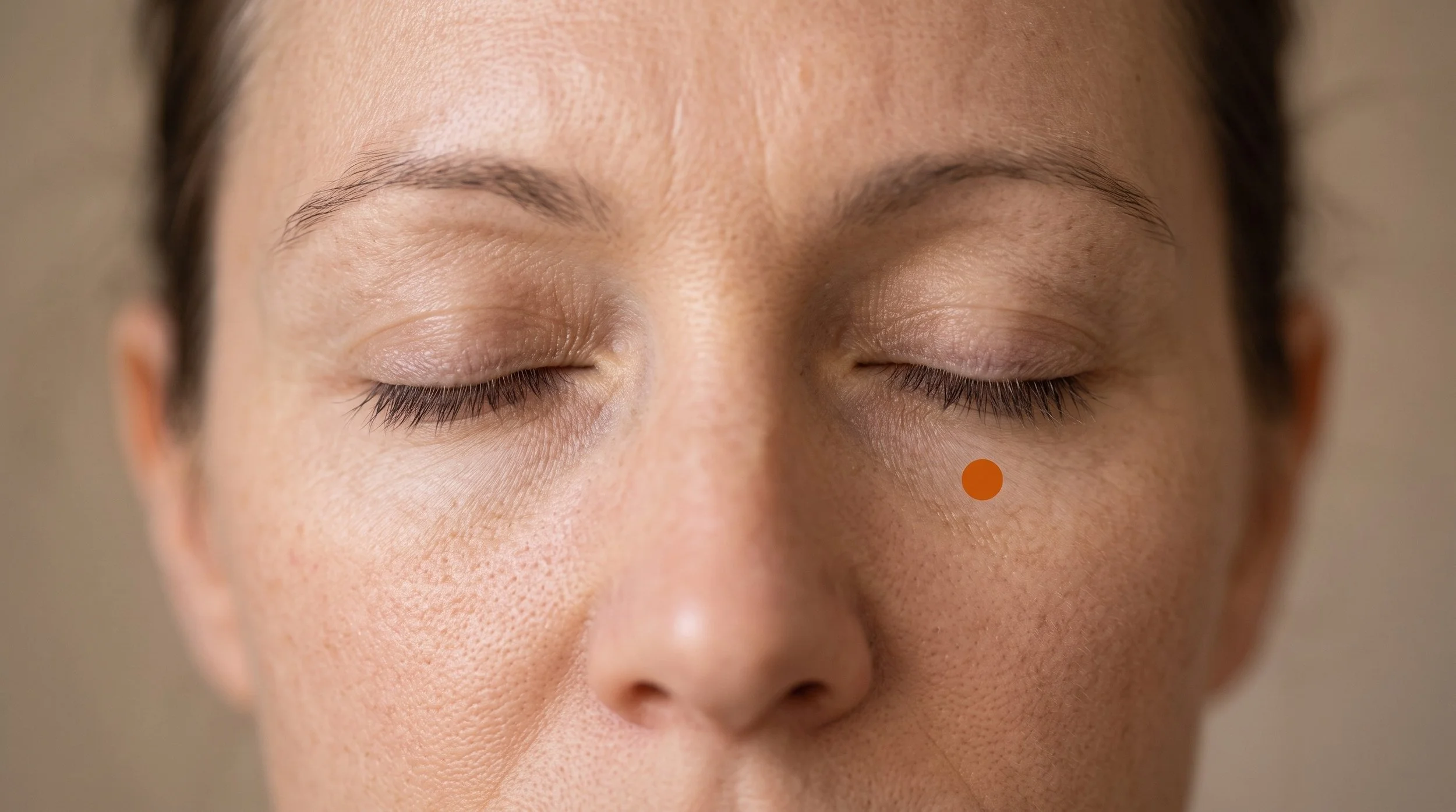
ST1, known in Chinese as Chengqi and usually translated as Container of Tears, is the first point of the Stomach channel and sits in one of the most delicate places on the body: directly below the pupil, in the narrow gap between the lower edge of the eyeball and the bony rim of the eye socket. Stomach 1 belongs to a very small club, since only a handful of classical points sit inside the orbital margin, and along with BL1 it is one of the two primary channel points that classical texts treat as the main local access to the eye itself.
That position is exactly why the st1 acupuncture point matters clinically and why it demands so much respect: the tissue here is thin, vascular, and richly innervated by trigeminal and facial nerve branches that regulate tearing, blinking, and eyelid tone, and the same features that make the container of tears point useful for red, watery, tired eyes also make it a point that belongs only in trained, supervised hands.
- ST1 lies directly below the pupil with the eyes looking straight ahead, in the sulcus between the eyeball and the infraorbital ridge, passing through the thinnest skin on the body, the palpebral fibers of orbicularis oculi, and the orbital septum before reaching pre-septal fat, with the bony orbital floor immediately below.
- Classical sources present ST1 as a meeting point of the Stomach channel with the Yang Motility and Conception vessels and credit it with brightening the eyes, stopping excessive tearing, and dispelling wind and heat from the eye region, traditional attributions that map onto modern interest in trigeminal, autonomic, and eyelid motor function rather than any literal flow of energy.
- The mechanism story here is a nerve story: the lower lid is supplied by infraorbital branches of the maxillary trigeminal division, the muscle by zygomatic branches of the facial nerve, and trigeminal afferents converge in the trigeminal spinal nucleus, a relay continuous with the upper cervical dorsal horn that also drives descending inhibitory and autonomic reflexes (Zhao, 2008).
- Point-specific physiology is limited but interesting: a randomized study in open-angle glaucoma found that an eye-specific needling scheme raised pulsatile ocular blood flow measurably while retinal flow, vessel diameter, and intraocular pressure did not change significantly (Leszczynska et al., 2018).
- The clinical research picture is mixed and cautious. ST1 appears in the periocular point sets used in randomized dry eye trials (Shin et al., 2010; Kim et al., 2012), while Cochrane reviews of acupuncture for childhood myopia, glaucoma, and Bell's palsy all concluded that trial quality was too limited for firm conclusions (Wei et al., 2011; Law et al., 2020; Chen et al., 2010).
- De qi at ST1 is deliberately understated. Practitioners do not lift, thrust, or rotate here, so patients typically report a light pressure or fullness under the eye rather than the spreading ache of a limb point, and firm pressure is applied for a full minute on withdrawal because bruising is the most common adverse effect of periocular needling (Lee et al., 2023).
Tired, Watery, Screen-Strained Eyes That Never Fully Settle?
Long hours at a monitor drop your blink rate, load the muscles around the orbit, and leave the eyes feeling dry, gritty, and heavy by mid afternoon. At Morningside Acupuncture we treat that pattern with a full head and neck approach, combining safe periorbital points, suboccipital and upper trapezius work, and distal points such as LI4 and LV3 rather than relying on any single spot. When periocular needling is appropriate, we use the guarded orbital technique with slow insertion and no manipulation. Book a visit and we'll build a plan around how your eyes actually behave through your workday.
Schedule NowAnatomy of ST1: Why the Infraorbital Rim Is Such an Important Location
The tissue at ST1 is unusually thin and unusually mobile. Skin over the lower eyelid is the thinnest on the body, and directly beneath it sit the palpebral fibers of orbicularis oculi, the sphincter-like muscle that closes the eye. Under that muscle lies a loose areolar plane, then the orbital septum and its pre-septal fat, and below the whole arrangement is the bony infraorbital rim. The eyeball rests immediately above, cushioned by orbital fat.
What loads this region day to day is mundane and relentless: reduced blink rate during screen work, squinting in bright light or with uncorrected refractive error, allergic rubbing, contact lens wear, poor sleep, and the sustained low-grade contraction of orbicularis oculi that many people carry as part of a habitual facial holding pattern.
The nerve geography explains most of what this point can and cannot do. Sensation over the lower lid and cheek travels in the infraorbital branches of the maxillary division of the trigeminal nerve, which exits the infraorbital foramen a short distance below ST1 at the level of ST2, while the conjunctiva and upper orbital structures are served by ophthalmic division branches. Motor supply to orbicularis oculi comes from zygomatic branches of the facial nerve.
All of the sensory traffic converges in the trigeminal spinal nucleus, a column continuous with the upper cervical dorsal horn, which is why eye-region symptoms, temple pain, and upper neck tension so often travel together and why needling in one territory may influence another through segmental convergence and descending inhibitory pathways (Zhao, 2008).
Tear production adds an autonomic layer: lacrimal secretion is driven by parasympathetic fibers reaching the gland by way of the pterygopalatine ganglion, with sympathetic input controlling orbital vascular tone, and both are reflexively linked to trigeminal input from the ocular surface. Studies suggest that eye-directed needling may shift ocular perfusion in the short term (Leszczynska et al., 2018).
Deeper still, the region carries infraorbital vessels, the fine vascular plexus of the eyelid, and orbital fat with the inferior oblique muscle in the vicinity, and the orbital floor itself is thin bone. This is why the needling conventions for ST1 are so specific and so conservative.
Classical instruction has the patient close the eyes and look upward while the practitioner gently guards the globe upward with a finger, then advances the needle slowly along the orbital floor to roughly 0.5 to 1 cun with no lifting, thrusting, or rotating, followed by firm pressure with a cotton ball for about a minute to limit hematoma. Moxibustion is classically contraindicated here.
A published review of severe periocular adverse events identified case reports of penetrating and perforating globe injuries, retrobulbar hemorrhage, and hematoma following eye-area needling, which is exactly why this technique belongs to supervised professional practice and why many clinicians substitute ST2 or BL2 when the clinical goal can be met outside the orbit (Lee et al., 2023).
Related Orbicularis Oculi Trigger Points Related Stomach ChannelST1 at a Glance: Classification, Location, and Clinical Use
| Category | Detail |
|---|---|
| Traditional Name | Chengqi, commonly translated as Container of Tears or Containing Tears; Stomach 1, the opening point of the Stomach channel |
| Channel Classification | First point of the Stomach channel of Foot Yangming, on the face at the lower orbital margin |
| Point Categories | Meeting point of the Stomach channel with the Yang Motility Vessel (Yang Qiao Mai) and the Conception Vessel (Ren Mai); one of the two principal classical local eye points of the primary channels alongside BL1; a convergence site where the Stomach sinew channel joins the Bladder channel sinew network around the eye and where the Stomach divergent channel connects with the eye |
| Precise Location | With the eyes looking straight ahead, directly below the pupil in the groove between the eyeball and the infraorbital ridge |
| Tissue Stimulated | Thin lower eyelid skin, palpebral fibers of orbicularis oculi, orbital septum and pre-septal fat, with the infraorbital rim as the bony floor and infraorbital nerve branches emerging just below |
| Needle Depth / Direction | Specialist orbital technique only: eyes closed and looking up, globe gently guarded upward, needle advanced slowly first slightly inferiorly then along the orbital floor to 0.5 to 1 cun, with no lifting, thrusting, or rotation, and firm pressure for about a minute on withdrawal; moxibustion is classically contraindicated |
| De Qi Sensation | Intentionally minimal, since the needle is not manipulated: usually a light pressure, fullness, or coolness under the eye, sometimes brief watering, without the spreading grasp typical of limb points |
| Primary Clinical Uses | Traditionally used for red, aching, itching eyes, tearing on exposure to wind, eye strain and dimness of vision, eyelid twitching, and facial paralysis involving the lower eyelid |
| Common Point Combinations |
|
Deadman's point compendium treats ST1 as one of the two great local eye points of the primary channels, and the reasoning it records is worth understanding as a traditional attribution rather than a physiological claim. In that framework, eye disorders were sorted into problems arriving from outside (wind with heat or cold), problems arising from within (patterns involving Liver fire, rising yang, or depleted Liver blood and yin), and mixtures of the two, and ST1 was considered useful across the whole range because it was thought to both expel external factors from the eye and strongly move local qi and blood.
The classical indication list is broad: redness, swelling and pain, excessive tearing, itching, dimness and short-sightedness, night blindness, visual dizziness, superficial obstruction of vision, lid twitching, and deviation of the mouth and eye. The name is the tell. Tearing was subdivided into cold and hot types, both complicated by wind, and a point named Container of Tears was placed precisely where tears pool.
What's striking is how closely the classical picture tracks the anatomy: a point named for tears, sited at the drainage corner of the lid, credited with governing lacrimation and lid movement, in the exact territory where trigeminal sensory and facial motor nerves meet.
Why ST1 Is Used for Eyelid Twitching, Wind-Induced Tearing, and the Facial Palsy Eye
The most useful way to think about ST1 is as a point sitting on top of a reflex loop. The surface of the eye and the lower lid report to the trigeminal system, the lid muscle takes orders from the facial nerve, and tear production is controlled by parasympathetic fibers that are themselves triggered by trigeminal input. Symptoms like watering in cold wind, a lid that flickers for weeks, or a lid that will not close properly after a facial palsy are all disturbances within that loop rather than diseases of the eyeball.
Needling in the same segmental territory feeds strong sensory input into the trigeminal spinal nucleus, where afferents from the face and the upper cervical spine converge, which is one plausible route by which local needling can change how a region feels and behaves (Zhao, 2008).
Beyond the local segment, needling recruits descending inhibitory pathways from the brainstem that dampen the transmission of nociceptive signals, and the large individual patient data analyses of acupuncture for chronic pain show effects that persist beyond the treatment session rather than fading immediately, which supports the idea that something more than short-term distraction is at work (Vickers et al., 2018). Autonomic effects are the other half of the story.
Vascular tone in the orbit is under sympathetic control, and a randomized study in glaucoma patients found that an eye-specific needling scheme produced a measurable short-term rise in pulsatile ocular blood flow while other parameters stayed flat (Leszczynska et al., 2018). That is a modest, single-study finding, not proof of a therapeutic effect, but it does suggest that periocular needling may reach the vascular and autonomic machinery around the eye.
In practice this means we set expectations honestly. ST1 may reduce the ache, heaviness, and irritability of a strained eye region and may help calm a twitching lid, and it's traditionally used for tearing, but no responsible practitioner should suggest that needling changes refractive error, glaucoma progression, or structural eye disease. The clinical value of the point is comfort, muscle tone, and local circulation, delivered as part of a broader treatment that usually includes the neck and distal points.
What the Research Shows for ST1
Almost all of the trial evidence relevant to ST1 comes from multi-point protocols, so no study isolates what this single point contributes. Where ST1 appears by name, it's one of a dozen or more periocular and distal points in dry eye protocols, which means the honest reading is that ST1 is part of point sets that have been tested rather than a point with its own evidence base. Reviews across ophthalmic indications also share a common limitation: many trials are small, conducted in single regions, and rated at high risk of bias, so most conclusions land somewhere between promising and inconclusive. The safety literature, by contrast, is quite specific and deserves as much attention as the efficacy literature.
| Study | Type | Focus | Key Finding |
|---|---|---|---|
| Lee et al., 2011 | Systematic review and meta-analysis | Acupuncture for dry eye compared with conventional care | Pooled results favored acupuncture on tear film measures, though the authors stressed that the small number of trials and their risk of bias limit confidence. |
| Shin et al., 2010 | Randomized sham-controlled trial | Moderate to severe dry eye using a 17-point protocol that included ST1 | Symptom and tear film measures improved within the treated group, but differences against sham needling were not clearly established. |
| Kim et al., 2012 | Multicenter randomized controlled trial | 150 patients with dry eye, acupuncture protocol including ST1 versus artificial tears | No statistically significant difference between groups on the primary symptom index, suggesting effects comparable to standard drops rather than superior to them. |
| Leszczynska et al., 2018 | Randomized clinical study | Short-term ocular blood flow in primary open-angle glaucoma | Pulsatile ocular blood flow increased after eye-specific needling, while retinal flow, vessel diameter, and intraocular pressure did not change significantly. |
| Wei et al., 2011 | Cochrane systematic review | Acupuncture for slowing myopia progression in children and adolescents | The available trials were too small and methodologically weak to show whether acupuncture slows myopia progression. |
| Law et al., 2020 | Cochrane systematic review | Acupuncture for glaucoma | The reviewers concluded that current data do not allow reliable conclusions about acupuncture as a treatment for glaucoma. |
| Chen et al., 2010 | Cochrane systematic review | Acupuncture for Bell's palsy, relevant to lower eyelid involvement | Six trials met inclusion criteria but design and reporting flaws prevented conclusions about efficacy, with no harms reported. |
| Lee et al., 2023 | Review of published case reports | Severe adverse events following periocular acupuncture | Fourteen case reports of serious ocular injury were identified over four decades, most involving globe penetration, underlining that orbital points require expert technique. |
Eyelid Twitching or Facial Weakness Around the Lower Lid?
Persistent lid flicker and post-palsy weakness both involve the orbicularis oculi and its facial nerve supply, and both respond best to consistent, well-sequenced care rather than one-off treatment. Our licensed acupuncturists assess facial muscle tone, cervical contribution, and sleep and stress load before choosing points, and we frequently work with ST2, BL2, GB20, and gentle facial needling instead of entering the orbit. If eye-area needling is indicated, it's performed only with the specialist guarded technique and clear consent. Schedule an evaluation to get a specific, staged plan.
Schedule NowST1 in the Context of Trigger Point Work
Acupuncture, in our definition, is the use of an acupuncture needle, and that includes the hundreds of styles practiced worldwide as well as dry needling. At Morningside we rarely approach the eye region through the orbit alone. Orbicularis oculi can develop taut, irritable bands that contribute to a tight, squinting, heavy-lidded feeling, and the trigger point literature describes referral from this muscle into the region above the upper lip and the side of the nose (Simons et al., 1999).
Those bands sit in the same territory ST1 addresses, but they can usually be treated with very superficial needling of the muscle belly outside the orbital margin, which achieves the muscular goal without any of the risk that comes with entering the orbit.
A typical session for eye strain and facial tension therefore layers techniques rather than relying on a single point. We commonly combine gentle facial needling near ST1 or ST2 with dry needling of temporalis, masseter, corrugator and procerus where indicated, plus the suboccipitals, upper trapezius, and levator scapulae, since upper cervical input converges with trigeminal input in the same brainstem relay. Distal points such as LI4 and LV3 round out the treatment.
The clinical rationale is simple: reduce the muscular load around the orbit, quiet the segmental input from the neck, and reserve true orbital needling for the cases where it clearly adds value and the patient has given informed consent.
ST1 Eye and Facial Treatment at NYC's Highest-Rated Acupuncture Clinic
Morningside Acupuncture is the highest-rated acupuncture and dry needling clinic in New York City with over 500 five-star Google reviews, and our team is trained in both classical channel theory and modern pain science. That combination matters for a point like Stomach 1, where knowing the anatomy of the orbital floor is as important as knowing the classical indications. We'll tell you plainly when ST1 is the right choice and when a safer neighboring point will do the same job. Schedule your appointment and let's get your eyes and face more comfortable.
Schedule NowFrequently Asked Questions
What does ST1 feel like when needled?
Much less dramatic than most people expect. Because the needle is never lifted, thrust, or rotated at this point, there's no attempt to produce the strong grasping ache associated with limb points. Most patients describe a brief pinprick as the needle passes the skin, then a mild pressure, coolness, or fullness under the eye, and sometimes a reflex watering of the eye that settles within seconds. The needle is usually retained quietly, and the practitioner presses the site firmly for about a minute afterward, which feels reassuring rather than uncomfortable. A small amount of bruising can appear over the following day even with careful technique, since eyelid tissue is loose and richly vascular.
Why needle the eye at all instead of using distal points on the hand and foot?
Often we do exactly that, and classical prescriptions for eye complaints almost always pair local points with distal ones such as LI4, LV3, LV2, or GB37. The argument for a local point is segmental: needling within the same trigeminal territory as the symptom delivers sensory input directly into the relay where those afferents converge, and it can influence local circulation and muscle tone in a way that a point on the hand cannot (Zhao, 2008). The argument against it is risk. Because ST2, BL2, and taiyang sit just outside the orbital margin and share much of the same innervation, many practitioners reach for those first and reserve ST1 for cases where the orbital position genuinely matters.
Can I press ST1 myself between sessions?
Yes, with an important boundary: press, never poke, and stay on the bone. Close your eyes, look straight ahead behind closed lids, and find the bony rim of your lower eye socket directly below the center of the pupil. Rest the pad of your index or middle finger on that bony ridge, not on the eyeball, and apply light, steady pressure aimed slightly downward into the bone for 20 to 30 seconds. Release, then repeat two or three times, and you can add small circular movements along the rim toward the outer corner. Two or three rounds a day, or once an hour during heavy screen work, is plenty. Keep the pressure gentle enough that it never feels sharp, stop immediately if you notice visual changes or pain, and skip it entirely if you have an eye infection, recent eye surgery, glaucoma under active management, or any undiagnosed change in vision. Pairing the pressure with a slow 20 second look at something far away is usually more useful than pressing harder.
Is ST1 safe to needle?
It's safe in trained hands and genuinely risky in untrained ones, which is why it's taught as a supervised technique. The eyeball sits directly above the needle path, the orbital floor is thin bone, and the surrounding tissue bruises easily. Standard precautions include eyes closed and looking up, the globe gently guarded upward, slow insertion along the orbital floor to no more than about 1 cun, no manipulation of the needle at any point, and firm pressure for a full minute on withdrawal. Moxibustion is classically contraindicated at this point. A published review of case reports documented serious injuries including globe penetration and retrobulbar hemorrhage after periocular needling, so this is not a point for self-treatment, for practitioners without specific orbital training, or for patients on anticoagulants without a careful risk discussion (Lee et al., 2023). Bruising remains the most common adverse effect even when everything is done correctly.
Where exactly is ST1 located?
Sit or lie with your eyes open and looking straight ahead. Draw an imaginary vertical line down from the center of your pupil. ST1 is where that line meets the space between the lower edge of the eyeball and the bony infraorbital ridge, roughly at the level of the lower lid margin. It's higher than most people guess: ST2 is the next point down, sitting in the small depression at the infraorbital foramen below the rim, and confusing the two is common. If your fingertip is resting comfortably on the cheekbone, you've gone too low.
References
- Deadman, P., Al-Khafaji, M., & Baker, K. (2009). A manual of acupuncture. Journal of Chinese Medicine Publications.
- White, A., Cummings, M., & Filshie, J. (2008). An introduction to Western medical acupuncture. Churchill Livingstone Elsevier.
- Simons, D. G., Travell, J. G., & Simons, L. S. (1999). Travell & Simons' myofascial pain and dysfunction: The trigger point manual, Vol. 1: Upper half of body (2nd ed.). Williams & Wilkins.
- Zhao, Z. Q. (2008). Neural mechanism underlying acupuncture analgesia. Progress in Neurobiology, 85(4), 355-375. https://doi.org/10.1016/j.pneurobio.2008.05.004
- Vickers, A. J., Vertosick, E. A., Lewith, G., MacPherson, H., Foster, N. E., Sherman, K. J., Irnich, D., Witt, C. M., & Linde, K. (2018). Acupuncture for chronic pain: Update of an individual patient data meta-analysis. Journal of Pain, 19(5), 455-474. https://doi.org/10.1016/j.jpain.2017.11.005
- Lee, M. S., Shin, B. C., Choi, T. Y., & Ernst, E. (2011). Acupuncture for treating dry eye: a systematic review. Acta Ophthalmologica, 89(2), 101-106. https://doi.org/10.1111/j.1755-3768.2009.01855.x
- Shin, M. S., Kim, J. I., Lee, M. S., Kim, K. H., Choi, J. Y., Kang, K. W., Jung, S. Y., Kim, A. R., & Kim, T. H. (2010). Acupuncture for treating dry eye: a randomized placebo-controlled trial. Acta Ophthalmologica, 88(8), e328-e333. https://doi.org/10.1111/j.1755-3768.2010.02027.x
- Kim, T. H., Kang, J. W., Kim, K. H., Kang, K. W., Shin, M. S., et al. (2012). Acupuncture for the treatment of dry eye: a multicenter randomised controlled trial with active comparison intervention (artificial teardrops). PLOS ONE, 7(5), e36638. https://doi.org/10.1371/journal.pone.0036638
- Leszczynska, A., Ramm, L., Spoerl, E., Pillunat, L. E., & Terai, N. (2018). The short-term effect of acupuncture on different ocular blood flow parameters in patients with primary open-angle glaucoma: a randomized, clinical study. Clinical Ophthalmology, 12, 1285-1291. https://doi.org/10.2147/OPTH.S170396
- Wei, M. L., Liu, J. P., Li, N., & Liu, M. (2011). Acupuncture for slowing the progression of myopia in children and adolescents. Cochrane Database of Systematic Reviews, 2011(9), CD007842. https://doi.org/10.1002/14651858.CD007842.pub2
- Law, S. K., Wang, L., & Li, T. (2020). Acupuncture for glaucoma. Cochrane Database of Systematic Reviews, 2020(2), CD006030. https://doi.org/10.1002/14651858.CD006030.pub4
- Chen, N., Zhou, M., He, L., Zhou, D., & Li, N. (2010). Acupuncture for Bell's palsy. Cochrane Database of Systematic Reviews, 2010(8), CD002914. https://doi.org/10.1002/14651858.CD002914.pub5
- Lee, S. M., Wu, J., & Hwang, D. D. J. (2023). Severe adverse events of periocular acupuncture: a review of cases. Korean Journal of Ophthalmology, 37(3), 255-265. https://doi.org/10.3341/kjo.2022.0111
Disclaimer: This web site is intended for educational and informational purposes only. Reading this website does not constitute providing medical advice or any professional services. This information should not be used for diagnosing or treating any health issue or disease. Those seeking medical advice should consult with a licensed physician. Seek the advice of a medical doctor or other qualified health professional for any medical condition. If you think you have a medical emergency, call 911 or go to the emergency room. No acupuncturist-patient relationship is created by reading this website or using the information. Morningside Acupuncture PLLC and its employees and contributors do not make any express or implied representations with respect to the information on this site or its use. For any legal interpretation of scope of practice in your state, consult a licensed attorney or regulatory authority.
Read more from our blog